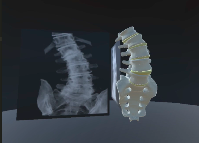
Medical images are taken in 3D space, getting information from coronal, axial, and sagittal views. This information can be translated into a 3D model; however, most programs require manual segmentation of separate anatomy parts for a anatomy separated model. In addition, these generated models are created as a volume rather than surface only.
Using the MLab in-house machine learning algorithm (Bilwaj Gaonkar, et al) on high resolution MRI (detailed in each plane), I am able to create a high quality 3D volumetric model using tools like Slicer 3D (). Using the Slicer SDK, this process can automatically generate an fbx model to import into any application.
As a separate process, I used the computer vision library OpenCV on Python language in order to also semi-automate the process. The process finds finds the center, angle, and dimensions of each vertebral body. This information is then fed into a Maya MEL script to automatically morph a generic 3D model into the patient-specific measurements. Comparisons can be seen on the bottom panel.
This is a work in progress, where future work will focus on increasing accuracy especially with pathologies present.